Positive staining TEM: Defined structures
Achieve an in-depth visualisation of the ultrastructural properties of your mammalian, insect or plant cell line. Ideally suited to screen your cells for viral or bacterial contaminations or to analyse complex intracellular processes in detail.
psTEM:
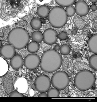
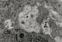
To visualise the morphology of your sample, your sample will be preserved by chemical fixation and then embedded in resin. The embedded sample will then be cut into ultrathin sections of about 50-70nm and then stained using heavy metal salts. The cellular structures of your sample will then appear dark on a bright background during imaging.
psTEM is ideally suited to:
Our method of psTEM for the detection of adventitious agents in cell substrates utilised in the production of biologics
For the detection of retrovirus-like particles in cell substrate, the cells are embedded in TEM resin, cut into ultrathin sections and stained using heavy metal salts. The staining agents directly contrast the cellular structures and for this reason this method is referred to as positive staining TEM. The ultrathin sections of cells are screened for adventitious agents. If retrovirus-like particles are detected, these particles are classified in type-A or type-C particles. Type-A particles are immature particles and are found in the cytoplasm, while type-C particles assemble at and bud from the host cell plasma membrane. It is not possible to quantify retrovirus-like particles in ultrathin sections, as virus particles are not expressed evenly across the cell population. For this reason, the percentage of infected cells is reported.
Results and the corresponding Final Reports (CoA) are issued within 15 working days.